
Dr. Leroy Butler shares his approach to treating Achilles tendon ruptures, explaining when surgery may be recommended, when nonsurgical treatment is appropriate, and why tendon length matters for long-term strength and recovery.
The phone call I get three days later
Somebody pushed off hard. A pickup game, a church league, a tennis serve, a sprint to first base. They felt a pop in the back of the ankle and turned around to see who kicked them. Nobody was there.
By the time they reach me, they have usually been to an emergency room or an urgent care, they are in a boot or a splint, and they have spent two nights reading. What they read does not agree with itself. One page says surgery is essential for athletes. Another says recent research proves surgery is unnecessary. A third describes a long incision down the back of the heel and a complication rate that sounds alarming. They are scared, they are rushed, and they think a decision has to be made today.
I have been performing Achilles repairs for more than fifteen years, and I have used the minimally invasive, jig-assisted technique known as PARS — the Percutaneous Achilles Repair System — for over a decade, long before a well-publicized professional football injury made “minimally invasive Achilles surgery” a phrase patients recognized. Let me start with the two things I say in the room before anything else.
First: this is almost never an emergency measured in hours. You have time to be evaluated properly and to make an informed choice. Even at three weeks out, most of my patients still undergo a standard PARS repair.
Second: the real question is not surgery or no surgery. The real question is what happens to the length of your tendon — because a ruptured Achilles can heal beautifully and still end up too long, and a long Achilles is a weak Achilles.
That single idea drives most of what follows.
What actually tore, and why it matters
The Achilles is the largest and strongest tendon in the body, connecting the calf muscles to the heel bone. When it ruptures, the two ends separate. The tendon is remarkably good at healing — it will scar across that gap whether or not anyone operates. The question is the distance it has to bridge, and where the ends settle while it does.
Think of it less like a broken bone and more like a rope under tension. If the ends knit back together with slack in the system, the rope still works, but you lose the ability to snap it tight. That is push-off power: the strength you need to climb stairs, jog, jump, and walk quickly.
This is not a theoretical concern. In a randomized trial of 60 patients from Finland comparing surgical and nonsurgical treatment with identical rehabilitation in both groups, tendons treated without surgery were on average 19 mm longer at 18 months, with more calf muscle atrophy and roughly 10–18% less plantarflexion strength (Heikkinen et al., AJSM, 2017). I should be fair about the strength of that evidence: it is one modest trial, and the American Orthopaedic Foot & Ankle Society’s 2024 review concluded there is not yet conclusive evidence that surgery prevents tendon elongation. It is the mechanism I find most persuasive, not a settled fact.
And here is the part that often gets left out: repair does not eliminate this. Long-term follow-up of surgically repaired patients found tendons still about 12 mm longer than the uninjured side, with 12–18% plantarflexion strength deficits fourteen years later (Heikkinen et al., JBJS, 2017). Surgery narrows the gap. It does not close it. Anyone who tells you an operation will make your Achilles exactly what it was is overselling.
This injury is also becoming more common. Incidence has been rising roughly 2.7% a year; it affects men about three times as often as women and peaks in men aged 30 to 49 (Kotsifaki et al., Sports Medicine, 2026). In U.S. data, basketball is the most common activity involved (Lemme et al., OJSM, 2018).
How I make the diagnosis — and why you probably don’t need an MRI
Most acute Achilles ruptures are diagnosed in the office, with hands and a history. Two findings do most of the work: a palpable defect, a gap I can feel in the tendon, and a positive Thompson test, in which squeezing the calf fails to move the foot because the tendon is no longer connected to it.
For a straightforward acute rupture, I usually do not order an MRI. I reserve advanced imaging for a tendon with pre-existing Achilles problems, or a rupture that is subacute or chronic, where I need to know how much viable tendon remains and how far the ends have retracted. A scan that will not change the plan mostly costs you days at the front end of a recovery that does better when it starts sooner.
What does change my plan is the pattern and the timing. A mid-substance tear behaves differently from a tear at the insertion into the heel bone, which is a different operation with a more extensive anchor construct. Time matters too, though less dramatically than patients fear. Scar tissue accumulates in the gap. At three weeks, most patients still get a standard PARS repair. Later than that, I may need a slightly larger incision to clear scar tissue first. Genuinely chronic ruptures may not be candidates at all, because the operation stops being a repair and becomes a reconstruction — tendon transfers, proximal lengthening. I perform those too, but I would rather not have to.
If there is one thing worth acting on today, it is this: get evaluated. Swedish data covering nearly a thousand ruptures found about 11% presented late enough to be considered chronic, and the large majority of that delay came from patients waiting to seek care rather than from clinicians missing the diagnosis (Nilsson et al., BMC Musculoskelet Disord, 2026). A chronic rupture is a harder problem than an acute one, and I would rather see you early and tell you that you have options than see you late and have fewer to offer.
Boot or surgery: what the evidence says, and what it doesn’t
Some patients arrive having read that modern research shows nonoperative treatment does just as well. That reading is not wrong, and I want to be straightforward about it: the published evidence is solid. Here is what it shows.
Surgery reduces re-rupture. One large analysis put it at 2.3% operatively versus 3.9% nonoperatively, while surgical patients had roughly three times the rate of other complications — and, importantly, found no significant re-rupture difference in the studies that used accelerated functional rehabilitation (Ochen et al., BMJ, 2019). Pooling randomized trials alone, the gap looks wider: re-rupture near 9–10% without surgery versus about 3% with repair, but with complications of any kind in roughly 19% of surgical patients versus 6% of nonoperative ones (Fan et al., Front Surg, 2024). The largest trial in the field, from Norway, analyzed 526 patients randomized to nonoperative care, open repair, or minimally invasive repair and found no meaningful difference in patient-reported function at one year, with re-rupture at 6.2% nonoperatively versus 0.6% in both surgical groups (Myhrvold et al., NEJM, 2022).
Study | Re-rupture: boot | Re-rupture: surgery | Trade-off |
Ochen, BMJ 2019 | 3.9% | 2.3% | ~3× other complications with surgery; no re-rupture difference with accelerated rehab |
Fan, Front Surg 2024 (RCTs) | ~9–10% | ~3% | Any complication ~19% surgical vs. ~6% nonoperative |
Myhrvold, NEJM 2022 (526 patients) | 6.2% | 0.6% | No meaningful difference in patient-reported function at one year |
Figures as reported in each source; studies differ in design and rehabilitation protocols.
Those numbers do not point in one direction, and I do not think patients are served by pretending they do. Surgery buys a lower re-rupture rate and costs you a higher rate of everything else.
Two observations about that body of work that I think patients deserve.
One: essentially none of it is American. The fourteen randomized trials pooled in the most recent meta-analysis were conducted in Canada, Sweden, Norway, Finland, Denmark, the UK, New Zealand, the Netherlands, Germany, Spain, and Romania — not one in the United States, and most in single-payer or universal-coverage systems. In my view, that matters: the patients enrolled, and the rehabilitation available to them, may not match a forty-five-year-old in North Texas who plays three nights a week. It is a question of how well the findings transfer, not whether the science is sound. And in fairness, the same trend exists here regardless: U.S. operative management fell from 18.3% to 12.3% between 2010 and 2019 (Wilder et al., Foot Ankle Spec, 2023).
Two: I see a selected group of patients, and you should weigh my observations accordingly. A meaningful part of my practice is second opinions. People come to me months after nonoperative treatment saying they walk slower than their spouse on a sidewalk, that stairs going down are the hard ones, that jogging feels wrong. I do not see the patients for whom the boot worked well, because they are fine and never call anybody. My clinical impression is real, and it is also biased. Both things are true.
So where do I land? I recommend repair for active people, and my reasoning is about power rather than re-rupture. If you want to advance through physical therapy more quickly and you want to protect push-off strength for athletics or a physically demanding job, repair is the option I recommend. I treat nonoperatively patients whose physiology argues against surgery: advanced physiologic age, significant medical comorbidities, a largely sedentary lifestyle. Both are legitimate paths — which is also the position of the American Orthopaedic Foot & Ankle Society, whose 2024 statement concluded that nonoperative treatment with early functional rehabilitation is effective and appropriate for select patients, and that surgical repair yields good outcomes with acceptable risk in healthy, active patients (AOFAS Position Statement, 2024).
When someone in their forties arrives determined to avoid surgery, I do not argue. I go back through the risks and benefits of both paths, ask specifically what activities they want to return to, and then I show them the whole decision tree — including what the operation looks like if this becomes a chronic rupture treated later, compared with a primary PARS repair now. Not to pressure anyone. Because you should be able to see all the doors before you pick one.
What a PARS repair actually is
A PARS repair is a minimally invasive Achilles repair performed through a small incision, usually 2 to 2.5 centimeters, just large enough to expose the upper end of the ruptured tendon.
A jig is inserted alongside the tendon, inside the paratenon — the sheath that surrounds it. The jig guides several sutures percutaneously through the tendon in a controlled plane, and those sutures are locked into the substance of the tendon. Through two additional small incisions near the heel, the sutures are passed down and secured with anchors in the calcaneus, creating a tendon-to-bone construct.
Why that construct, rather than simply tying the two tendon ends back together? Two reasons, and they are really the same reason. Both are matters of surgical judgment rather than settled evidence, and I’ll label them as mine.
I try not to leave suture knots inside the rupture zone. My concern is that knots sitting in healing tissue can settle, and settling means lengthening.
I believe anchoring into bone gives me a stronger construct with better resistance to elongation. That confidence is what allows me to advance a patient through therapy sooner. It comes back to the same principle:
Protect the length, protect the power.
The construct is chosen by the rupture, not the other way around. Some patterns are best served by end-to-end repair. Tears at the insertion get a different operation entirely, with a more extensive anchor construct. No single technique is right for every tendon.
The operation takes about thirty minutes in my hands. It is outpatient and typically done under general anesthesia — you go home the same day, in a splint.
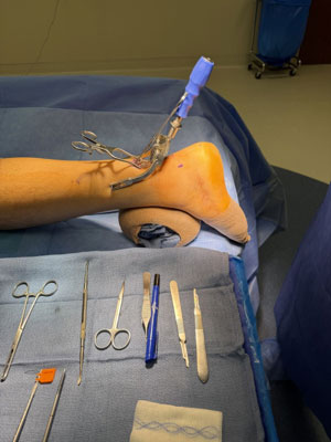
Intraoperative view of a minimally invasive Achilles repair. The working incision above the heel is small enough that a single self-retaining retractor spans it, and the instruments laid out on the tray are the entire set the operation requires. Photograph published with patient authorization.
What the jig is for, and what patients ask about the nerve
The question I get most often about this operation is whether a smaller incision means something got done blind. It’s a fair question, and the answer is the jig.
The jig exists to control where the sutures travel. That matters because of the sural nerve, which supplies sensation along the outer border of the foot and runs through territory percutaneous sutures have to cross. Freehand percutaneous techniques pass those sutures without direct control, and nerve injury has been a real and recognized problem with them — pooled data put it near 5.6%. Jig-assisted repair brings that to roughly 0.57%, because the sutures are guided inside the paratenon rather than passed through tissue no one can see (Melinte et al., Int Orthop, 2025). A cadaveric study of the PARS jig specifically found no nerve punctures across 50 needle passes, while also documenting that the working margin is a matter of millimeters (McGee et al., Foot Ankle Surg, 2021). It is a technique that rewards care rather than one that removes risk.
So this is a safe operation with a low absolute rate of nerve problems, and the jig is the reason. In my own practice, across several hundred of these repairs, I can account for about three patients with sural nerve irritation I would attribute to the surgery — and all of them resolved on their own with time. That is my experience rather than a published series, and I’d rather give you the honest number than a rounder one.
Worth separating out, because patients conflate the two: some people arrive with numbness or tingling along the outside of the foot from the rupture itself, before anyone has operated. If you already have those symptoms, tell your surgeon before surgery — it changes how we interpret anything you feel afterward.
What I won’t tell you is that a minimally invasive repair beats a well-performed open repair on nerve safety. The published comparisons don’t show that, and in some randomized data nerve irritation is reported more often after minimally invasive surgery. Open repair remains a good operation, and I still use open techniques when the rupture pattern calls for it.
I chose the smaller approach for the skin, not for the nerve. That is where the difference is real. Compared with traditional open repair, minimally invasive techniques consistently reduce wound complications and superficial infection: one meta-analysis of randomized trials found superficial infection in 6.0% of open repairs versus 0.4% of minimally invasive repairs (Attia et al., AJSM, 2023), and another found wound complications reduced by roughly 87% (Grassi et al., JBJS, 2018). In a retrospective series of 101 PARS repairs compared with 169 open repairs, 98% of patients returned to baseline activity by five months versus 82% after open repair (Hsu et al., FAI, 2015). Keeping the wound question in proportion, though: in a modern series of 369 consecutive open repairs, about 9% had some wound issue and only 0.54% needed another operation for it (Bishop et al., FAO, 2023).
My preference for the smaller approach came from experience, not from a brochure. The skin over the back of the heel is thin and poorly vascularized, and it does not forgive. I have treated wound complications from open Achilles repairs — my own patients’ and other surgeons’ — and while serious wound problems are uncommon, one in that location can turn a good operation into a bad year.
Who should not have this operation, or not right now
Publishing the reasons I say no is the fairest way to explain the reasons I say yes. I do not recommend Achilles repair for patients with:
- Moderate to severe vascular insufficiency
- Existing wound problems in the surgical area
- Poorly controlled diabetes
- Significant pre-existing neuropathy
- Active nicotine use — smoking or vaping — without a willingness to stop through surgery and recovery
Among more than 24,000 primary Achilles repairs, diabetes and vascular disease were associated with the highest surgical site infection rates (Dombrowski et al., KSSTA, 2019).
Nicotine is its own category. I verify with a preoperative nicotine lab test rather than relying on the conversation. This is not moralizing — it is arithmetic. In a matched analysis of more than 4,000 Achilles repairs, nicotine dependence was associated with roughly 55% higher wound disruption and 64% higher infection risk (DeShazo et al., FAI, 2024).
Sometimes I delay rather than decline, and I tell those patients what delay costs: waiting may mean the repair can no longer be done through as small an incision. And sometimes I talk people out of surgery altogether. Some patients are better served by a boot and an honest conversation.
Recovery: what it actually looks like
Nothing below is a guarantee. Recovery varies with the patient, the rupture, and the rehabilitation available to them. These are typical ranges in my practice.
FIRST TWO WEEKS
Splint, then transition to a boot with a heel lift at the first postoperative visit.
TWO TO FOUR WEEKS
Begin weight-bearing in the boot, timed to how the incision looks and how you are doing. Before that, you will be non-weight-bearing on that leg and will need crutches, a walker, or a knee scooter.
DESK WORK
Often within several days. A heavy-labor job is a different conversation and may wait until the boot comes off.
DRIVING
Typically possible around four weeks if the injury is on the left side and you drive an automatic. A right-sided repair usually takes six to eight weeks. In either case you must have reasonable ankle control, be off sedating medication, and remove the boot to drive.
AROUND EIGHT WEEKS
Begin dropping down from the heel lift with your physical therapist, toward flat in the boot.
TEN TO TWELVE WEEKS
Out of the boot and into a normal shoe for the average patient. Higher-level athletes sometimes progress faster.
THREE TO FIVE MONTHS
Most patients are jogging.
CLEARED FOR CUTTING AND JUMPING
Once you can perform a single-leg heel rise on the operated side.
That last one is the most important line in this section. The calendar does not clear you. Your heel rise clears you. It is why two people who had surgery the same week get cleared months apart, and it puts the milestone where it belongs — in your control.
Two things patients do not expect, so I tell them in advance.
You will look recovered before you are recovered. At ten to twelve weeks you are walking in a normal shoe with very little power in the tendon or calf. You will walk more slowly. The ankle will swell. That gap between looking fine and being strong is where people lose faith.
The heel gets more sore as you get more active, and that is the handoff. Early on, the anchors are doing work the tendon is not yet strong enough to do. As the tendon remodels and takes that job back, the area gets irritated. Patients expect pain to decrease steadily, so an uptick reads as failure. It usually is not. What frustrates my patients most is not the pain — it is how long the road back to all-out sport really is.
I define a successful outcome at nine to twelve months, when a patient is back to their previous level of activity. Published return-to-sport rates hover around 80%, though that figure falls when studies define return to play rigorously (Zellers et al., BJSM, 2016). Even among professional athletes, about 76% return, at a mean of eleven months (Johns et al., FAI, 2021).
Which brings me to the expectation I most often have to reset. Since a high-profile professional football Achilles repair and rapid comeback made headlines, patients arrive expecting that timeline. He rehabilitated for hours a day with a full-time medical staff built around getting him back. You have a job, a commute, and a family. Most people do return to their prior activities — just not on a professional’s schedule.
Whatever operation you have, what you cannot have is a professional athlete’s calendar.
I should also mention calf size honestly, because lifters ask and most articles avoid it: some calf atrophy is common after an Achilles rupture, and you may not get the same calf measurement back.
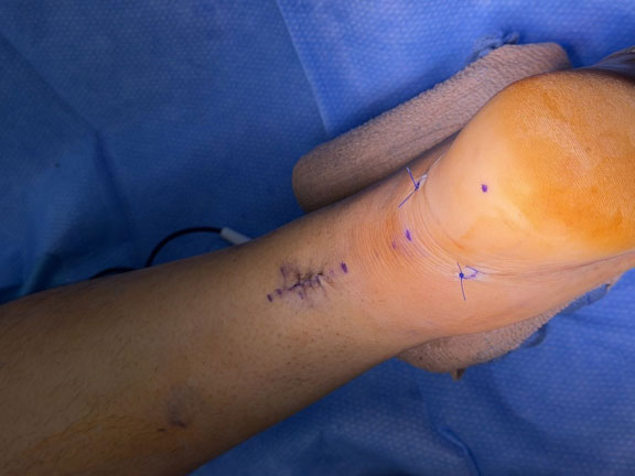
The same repair immediately after closure. The proximal incision — where the tendon ends are reached — measures roughly 2 to 2.5 centimeters. The two smaller incisions closer to the heel are where the sutures pass down and are secured with anchors in the calcaneus. This photograph shows incision size at the time of surgery, not healed appearance; scars continue to change for many months, and individual results vary. Published with patient authorization.
Experience, and one patient who changed how I practice
I perform approximately 30 to 40 mid-substance Achilles repairs a year, not counting insertional reconstructions — several hundred over roughly a decade in the Metroplex. I have been an orthopedic surgeon for more than fifteen years, practicing in North Texas for nearly ten. I am board certified in orthopedic surgery by the American Osteopathic Board of Orthopedic Surgery and fellowship-trained in foot and ankle surgery through the Las Vegas Foot and Ankle Fellowship, and I hold privileges at Legent Orthopedic Hospital, Legent Coppell Surgery Center, Eminent Medical Center, Texas Health Frisco, Medical City Frisco, Carrollton Regional Medical Center, and Sherman Medical Center. I also teach minimally invasive foot and ankle technique to other practicing surgeons through courses and cadaver labs, and I have been involved in implant and technique development.
Most of my Achilles patients come from Dallas–Fort Worth and the surrounding North Texas area, and many arrive by word of mouth — sent by a teammate or a friend who tore theirs and had it repaired. Patients also travel from outside Texas for complex, revision, and second-opinion foot and ankle care.
The case that changed my practice was a weekend athlete and serious lifter who found me online specifically because he had read about PARS repair. He did everything right on pain control and elevation, then pushed his weight-bearing and therapy considerably faster than I had asked him to. He did extremely well.
I am not recommending that anyone ignore their surgeon. But watching a patient outrun my protocol and finish strong made me systematically re-examine a rehabilitation timeline I had largely inherited. Since then I have advanced most of my patients faster and more deliberately — and it reinforced my confidence in taking the repair down to anchors, because a stronger construct is what makes earlier progression reasonable in the first place.
One patient’s result does not predict yours. But it is an honest account of how a surgeon’s thinking actually changes.
If you are dealing with a torn Achilles
If you heard a pop in the back of your ankle and you are having trouble pushing off, you should be evaluated by a foot and ankle specialist — whether or not you end up having surgery. A physical examination is necessary to make this diagnosis properly; a telehealth visit can begin the conversation and help organize records and imaging, but it cannot complete the evaluation.
Contact our team at the Orthopedic Institute of North Texas to schedule an evaluation, or speak with one of our representatives about the records, imaging, and next steps needed to begin the process. If you have already been treated elsewhere and are not getting your power back, a second opinion is reasonable.
You do not need to decide on surgery today. You only need to take the next informed step.
Frequently asked questions
How do I know if I tore my Achilles tendon or just sprained my ankle?
An Achilles rupture usually involves a sudden pop or snap in the back of the ankle during a push-off, often with the sensation of being kicked or struck from behind, followed by difficulty pushing off when walking. An ankle sprain typically involves rolling the ankle, with pain and swelling around the ankle joint itself. The diagnosis is confirmed on examination by feeling a gap in the tendon and by a Thompson test. If you are unsure, get examined — a delayed diagnosis is harder to treat.
Do I need an MRI to diagnose an Achilles rupture?
Usually no. Most acute Achilles ruptures are diagnosed clinically from the history and physical examination. I order an MRI selectively — for patients with pre-existing Achilles problems, or for subacute and chronic ruptures where I need to see how much tendon remains and how far the ends have retracted.
Can an Achilles rupture heal without surgery?
Yes. The tendon will heal whether or not it is repaired, and nonoperative treatment with early functional rehabilitation is appropriate for many patients. The trade-offs are a higher re-rupture rate and a tendon that tends to heal in a lengthened position, which is associated with more calf atrophy and less push-off strength. That trade matters most for people whose activities depend on power.
Is it too late for surgery if I tore it two or three weeks ago?
Usually not. At three weeks, most of my patients still undergo a standard PARS repair. Longer delays may require a slightly larger incision to clear scar tissue, and truly chronic ruptures may need reconstruction rather than repair. If you are within a few weeks of the injury, get evaluated rather than assuming you have missed your window.
Is nerve injury a risk with minimally invasive Achilles repair?
The sural nerve, which gives sensation to the outer border of the foot, is the nerve of concern. With freehand percutaneous techniques that pass sutures without direct control, injury has been reported in roughly 5–6% of cases. Jig-assisted repair, where the sutures are guided inside the tendon sheath, brings that to well under 1%. In my own practice, across several hundred repairs, about three patients have had sural nerve irritation I would attribute to surgery, and all resolved with time. Minimally invasive repair does not have a nerve-safety advantage over a well-performed open repair — its advantage is in wound healing. And some patients have numbness along the outside of the foot from the rupture itself, before any surgery; tell your surgeon if that describes you.
How big is the incision for a PARS Achilles repair?
Typically about 2 to 2.5 centimeters — roughly an inch — plus two small incisions near the heel where the anchors are placed. The operation takes about thirty minutes in my hands and is performed as an outpatient procedure.
When can I drive, return to work, and go back to sports after Achilles surgery?
In my practice, driving is often possible around four weeks for a left-sided repair in an automatic vehicle, provided you have adequate ankle control and are off sedating medication; a right-sided repair generally takes six to eight weeks. Desk work often resumes within several days; physically demanding work usually waits longer, sometimes until the boot comes off. Most patients are out of the boot at ten to twelve weeks, jogging between three and five months, and cleared for cutting and jumping once they can perform a single-leg heel rise. These are typical ranges, not promises.
Why does my heel hurt more as I get more active after surgery?
This is common in patients with anchors, and I warn people about it in advance. Early on, the anchors carry load the healing tendon cannot yet handle. As the tendon remodels and takes that work back, the area often becomes more irritated. Increasing soreness with increasing activity is usually part of that handoff rather than a sign something has failed — but new sudden pain, a pop, sudden weakness, fever, or drainage from the incision should be evaluated promptly.
Who is not a good candidate for Achilles repair surgery?
Patients with moderate to severe vascular insufficiency, active wound problems in the area, poorly controlled diabetes, significant neuropathy, or ongoing nicotine use who are unwilling to quit. The skin over the back of the heel heals poorly, and a wound complication there can lead to additional procedures. I test for nicotine before surgery, and I do sometimes recommend against operating.
Will my calf ever look and feel normal again?
Some calf atrophy is common after an Achilles rupture, and you may not regain your previous calf size. Measurable strength deficits can persist even after a successful repair. Most patients return to their prior activities; complete normalization is not something I promise.
I had my Achilles treated in a boot elsewhere and I still feel weak. Is a second opinion worth it?
Yes. If you notice you are walking slower than the people around you, struggling going down stairs, or unable to jog or jump the way you did, those are worth evaluating. Options depend on how much time has passed, tendon length, and calf strength — and not everyone needs another operation.
Do you see patients from outside Dallas–Fort Worth?
Most Achilles patients come from DFW and surrounding North Texas. Patients do travel from outside the region for complex, revision, and second-opinion foot and ankle care. A telehealth visit can begin that process, but an in-person examination is required to complete the evaluation.
A note on disclosure. I consult for several orthopedic device manufacturers, and I receive royalties on implants and techniques I helped develop. I mention it because you should know what financial relationships a surgeon has before you weigh my recommendation — and because none of the companies I work with make the system described in this article.
This article is for general education and is not medical advice or a substitute for an in-person evaluation. Individual results vary; no outcome or recovery timeline is guaranteed. Please consult a qualified physician about your specific situation.
Sources
- Attia AK, et al. Outcomes and complications of open versus minimally invasive repair of acute Achilles tendon ruptures. Am J Sports Med. 2023;51(3):825–836. doi
- Bishop ME, et al. Open repair of acute Achilles tendon ruptures: is the incidence of clinically significant wound complications overestimated? Foot Ankle Orthop. 2023;8(3). doi
- DeShazo S, et al. Nicotine dependence and rates of postoperative complications in Achilles tendon repair. Foot Ankle Int. 2024;45(1):67–72. doi
- Dombrowski M, et al. Medical comorbidities increase the rate of surgical site infection in primary Achilles tendon repair. KSSTA. 2019;27(9):2840–2851. doi
- Fan L, et al. Surgical vs. nonoperative treatment for acute Achilles’ tendon rupture: a meta-analysis of RCTs. Front Surg. 2024;11:1483584. link
- Grassi A, et al. Minimally invasive versus open repair for acute Achilles tendon rupture. J Bone Joint Surg Am. 2018;100(22):1969–1981. doi
- Heikkinen J, et al. Soleus atrophy is common after the nonsurgical treatment of acute Achilles tendon ruptures. Am J Sports Med. 2017;45(6):1395–1404. doi
- Heikkinen J, et al. Tendon length, calf muscle atrophy, and strength deficit after acute Achilles tendon rupture. J Bone Joint Surg Am. 2017;99(18):1509–1515. doi
- Hsu AR, et al. Clinical outcomes and complications of Percutaneous Achilles Repair System versus open technique. Foot Ankle Int. 2015;36(11):1279–1286. doi
- Johns W, et al. Career outlook and performance of professional athletes after Achilles tendon rupture. Foot Ankle Int. 2021;42(4):495–509. doi
- Kotsifaki R, et al. Incidence, temporal trends, and surgical shift of Achilles tendon rupture. Sports Med. 2026;56:1467–1487. doi
- Lemme NJ, et al. Epidemiology of Achilles tendon ruptures in the United States. Orthop J Sports Med. 2018;6(11). doi
- McGee R, et al. Anatomic relationship of the sural nerve when performing Achilles tendon repair using the Percutaneous Achilles Repair System. Foot Ankle Surg. 2021;27(4):427–431. doi
- Melinte MA, et al. Mini-open versus percutaneous surgical repair for acute Achilles tendon rupture. Int Orthop. 2025;49(1):259–269. doi
- Myhrvold SB, et al. Nonoperative or surgical treatment of acute Achilles’ tendon rupture. N Engl J Med. 2022;386(15):1409–1420. doi
- Nilsson N, et al. Incidence, reason for treatment delay and patient-reported outcome of patients affected by a chronic Achilles tendon rupture. BMC Musculoskelet Disord. 2026;27:371. doi
- Ochen Y, et al. Operative treatment versus nonoperative treatment of Achilles tendon ruptures. BMJ. 2019;364:k5120. link
- Wilder JH, et al. Rates of operative management for Achilles tendon rupture over the last decade. Foot Ankle Spec. 2023;16(4):377–383. doi
- Zellers JA, et al. Return to play post-Achilles tendon rupture: a systematic review and meta-analysis. Br J Sports Med. 2016;50(21):1325–1332. doi
- American Orthopaedic Foot & Ankle Society. Position Statement: Management of Acute Achilles Tendon Ruptures. November 2024. link

Leroy Butler, D.O., FAAOS.
Foot & Ankle Orthopedic Surgeon
Dr. Leroy Butler is a board-certified, fellowship-trained orthopedic foot and ankle surgeon at the Orthopedic Institute of North Texas, serving patients throughout the Dallas–Fort Worth area.
Dr. Butler specializes in minimally invasive Achilles tendon repair, total ankle replacement, minimally invasive bunion surgery, sports injuries, and complex foot and ankle reconstruction. He also has extensive experience treating ankle instability, arthritis, deformities, and challenging revision cases.
Beyond his clinical practice, Dr. Butler teaches advanced surgical techniques to other orthopedic surgeons and contributes to the development of orthopedic implants and surgical technologies.
He is committed to providing individualized, evidence-based care, considering both surgical and nonsurgical treatment options to help patients restore mobility, improve function, and return to the activities they enjoy.